Materials and methods: Morphological and morphometric study of trephine biopsy specimens of bone tissue was performed in three groups of patients 180 days after sinus lift surgery using different compositions of bone grafting material: Group 1 – Cerabone® granules (100%); Group 2 – Cerabone® with autograft bone chips in a 25:75 ratio; Group 3 – Cerabone® with autograft bone chips in a 50:50 ratio. Microscopic picture was assessed on sections stained with hematoxylin and eosin, volume fractions of the main components of the regenerate and cellular activity indices were measured.
Results: Direct osteogenesis with the formation of mature cancellous bone was observed in all groups. The highest volume of newly formed bone tissue (BV) was achieved in Group 3 (34.5%), while the lowest one was detected in Group 1 (25.75%). Indices of osteoblastic activity (N.ob, Ob.S) and material integration (MatBS/BS) were also highest in Group 3 (70.2%).
Conclusion: The results of our study indicate the advantage of using composite bone grafting materials with a 50:50 ratio of Cerabone® and autogenous bone graft for optimizing bone repair in sinus lift and emphasize the need for morphometric monitoring of the effectiveness of various bone graft compositions in reconstructive surgery.
Introduction
The success of dental implantation in the posterior maxilla directly depends on the availability of sufficient volume of bone tissue. Alveolar ridge atrophy due to prolonged tooth loss, pneumatization of the maxillary sinus, and some anatomical features often contribute to insufficient bone height for reliable implant anchorage [1]. To address this issue, sinus lift surgery is used in clinical practice. It increases bone volume by raising the floor of the maxillary sinus and filling the resulting space with graft material [2].
The choice of material for repairing bone defects in the sinus lift zone remains a crucial issue in contemporary dentistry [3, 4]. An ideal bone grafting material should provide three main functions: osteogenesis (that is, to supply progenitor cells), osteoinduction (i.e., to stimulate osteoblast differentiation), and osteoconduction (that is to serve as a matrix for new bone growth). Currently, bone autograft is considered the gold standard against which all other materials are compared, because it provides all above-mentioned functions and does not cause immunological rejection [5]. However, harvesting bone autograft requires additional surgical intervention, increases the operating time and risk of postoperative complications, while the volume of material available for harvesting is often quite limited [6, 7].
Consequently, alternative materials (allogeneic, xenogeneic, and synthetic) are under development and practical implementation [8, 9]. The xenogeneic material Cerabone® (Botiss Biomaterials GmbH, Germany) is granulated bovine hydroxyapatite, subjected to high-temperature processing to remove organic components. Consequently, it exhibits high biocompatibility, osteoconductive properties, and slow resorption, providing long-term preservation of the reconstructed bone volume [4]. However, just like other xenogeneic and synthetic materials, Cerabone®’s osteogenic and osteoinductive properties are somewhat limited, and the process of its replacement with bone autograft tissue is slower [10].
A promising approach to optimizing bone repair is the use of composite bone materials in which bone is combined with an osteoconductive material. It is believed that this grouping combines the advantages of both types of materials: bone autograft provides a supply of osteogenic cells and growth factors, while the granules of the xenogeneic material serve as a reliable scaffold, preventing excessive resorption and maintaining volume [11, 12, 13]. Many clinical and experimental studies confirmed the effectiveness of this approach [14-17]. Nevertheless, the question of the optimal ratio of autogenous bone to the xenogeneic component remains open. Comparative morphometric studies that allow quantitative assessment of the effect of the component ratio on the volume and quality of newly formed bone over the long term are rare, and their result are contradictory [18].
Quantitative assessment of bone regenerate using morphometric methods is essential for an unbiased comparison of different materials in terms of their effectiveness. Analysis of parameters such as the volume fraction of newly formed bone tissue (BV), the number of osteoblasts (N.ob, N.Mat.ob), and the level of integration of the implant material with the bone (MatBS/BS) allows assessing the current state of the regenerate, along with predicting its capability to withstand functional loads after subsequent implantation.
Currently, despite extensive research on sinus lift, sufficient data are lacking for a systematic comparative analysis. For instance, it was not studied yet how different proportions of bone autograft and Cerabone® granules affect the morphometric parameters of bone repair during the period corresponding to complete consolidation of the graft.
The objective of this study was to conduct a comparative morphometric analysis of bone repair processes in the sinus lift zone using various compositions of bone grafting materials.
Materials and methods
The study was performed on trephine biopsy specimens of bone tissue from patients who underwent sinus lift surgery using Cerabone® bone grafting material (approved for use in the Russian Federation: registration certificate No. FSZ 2011/09299) followed by dental implantation. Patients were distributed among three groups based on the bone grafting material composition. In Group 1 patients, the subantral space was filled with pure Cerabone® granules (100%). Group 2 individuals were subjected simultaneously to Cerabone® granules and autograft bone chips (taken from the maxillary tuberosity or mandibular ramus) in a 25:75 ratio. Group 3 patients received both components (Cerabone® and autograft bone chips) in equal amounts, i.e., in a 50:50 ratio. The sample size in each group was 10 patients.
The surgery was performed using a standard lateral approach to the maxillary sinus. Later, 180 days after the sinus lift, trephine biopsy specimens of bone tissues were collected from the area where the implants were to be placed under local anesthesia. The resulting bone tissue specimens were fixed in 10% neutral buffered formalin at room temperature for 48 hours. After fixation, the specimens were decalcified in 10% ethylenediaminetetraacetic acid (pH 7.2–7.4), and then soaked and embedded in paraffin.
Sections (5–7 µm thick) were cut from each paraffin block on a rotary microtome, followed by staining with hematoxylin and eosin according to the standard protocol.
Microscopic examination was performed using a LeicaDM2500 light microscope (Leica Microsystems GmbH, Germany) at magnifications of 100, 200, and 400.
Quantitative morphometric analysis was performed using Leica QWin image analysis software (Leica Microsystems GmbH, Germany). For each specimen, at least 10 non-overlapping fields of view were analyzed at a magnification of 200×. The following parameters were determined according to recommendations for bone tissue histomorphometry:
1) N.ob (cells/mm) – mean number of osteoblasts per 1 mm of the surface length of bone trabeculae and the surface of the bone grafting material;
2) N.Mf (cells/mm²) – mean number of macrophages in the stroma of the intertrabecular spaces per 1 mm² of section area;
3) N.Mat.ob (cells/mm) – mean number of osteoblasts in direct contact with the surface of the graft material granules per 1 mm of the material surface length;
4) BV (bone volume, %) – volume fraction of mineralized bone tissue (both mature and newly formed) of the total volume of the biopsy specimen;
5) FbV (fibrous tissue volume, %) – volume fraction of fibroreticular (connective) tissue;
6) MatV (material volume, %) – volume fraction of residual bone grafting material;
7) Ob.S (osteoblast surface) – specific bone surface area covered by active osteoblasts (the ratio of the length of the surface lined by osteoblasts to the total length of the bone surface);
8) Mat.Ob.S/Mat.S (%) – fraction of the material surface occupied by osteoblasts (the ratio of the length of contact of osteoblasts with the material to the total length of the material surface);
9) MatBS/BS (material-bone contact, %) – the relative area of the contact of newly formed bone with the graft material surface (the ratio of the length of areas where the bone is adjacent to the material to the total length of the graft material surface).
Volume fractions (BV, FbV, MatV) were measured via point counting using an Avtandilov test grid included in the microscope eyepiece, or automatically on digital images using morphometric software.
The study was approved by the Ethics Committee at the Central Research Institute of Dentistry and Maxillofacial Surgery of the Russian Federation Ministry of Healthcare. All patients signed informed consent to participate in the study.
Statistical processing of the collected data was performed using the Statistica 12.0 software package (StatSoft, USA). The arithmetic mean (M) and sample standard deviation (SD) were calculated for each group. Data are presented as M±SD. The normality of distribution was tested using the Shapiro-Wilk test. Intergroup differences were assessed using the Student’s t-test for independent samples (for normally distributed data) or the Mann-Whitney U test (in case of non-normal distribution). The data were considered statistically significant at p<0.05.
Results
Trephine biopsy specimens of bone tissue obtained 180 days after sinus lift surgery in three patient groups were examined under the microscope. Qualitative and quantitative morphological analyses of the structural components of the regenerated bone were conducted.
In Group 1, the material consisted predominantly of mature cancellous bone tissue, characterized by a pronounced trabecular structure with an orderly arrangement of bone trabeculae. Osteocytes possess a well-defined structure; they are located within the trabeculae and are enclosed in bone lacunae; the compact intercellular substance is uniformly stained. Loose connective tissue with capillaries, macrophages, lymphocytes, histiocytes, isolated eosinophils, and islets of lipocytes is visible in some of the intertrabecular spaces. In other spaces, layers of irregular dense connective tissue with arterioles and small arteries, individual macrophages, and lymphocytes are noticeable (Figure 1).
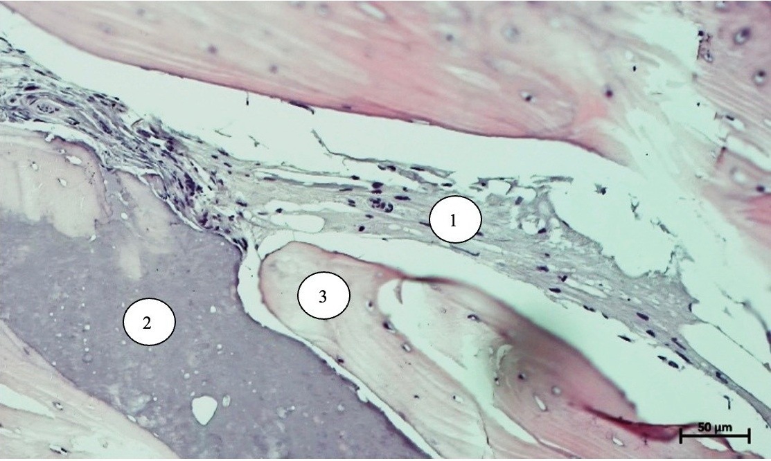
Figure 1. Loose connective tissue (1), osteoid with bone lamellae and bone grafting material (2), newly formed bone tissue (3). Hematoxylin and eosin staining; 200×.
Homogeneous eosinophilic deposits corresponding to osteoid are observed on the surface of some trabeculae, indicating ongoing bone formation. Islets of bone lamellae are visible within the osteoid. The structure reveals foci of bone grafting material (microgranular matrix) and fragments of bone trabeculae (bone autograft material) integrated into the surrounding tissue. Immature bone trabeculae are present as well; on the surface, they are covered by osteoblasts and have intensely stained intercellular substance. Group 1 is characterized by optimal combination of mature bone structures with zones of ongoing osteogenesis (Figure 2).

Figure 2. Newly formed bone tissue (1), osteoid with bone lamellae and bone grafting material (2), loose connective tissue (3). Hematoxylin and eosin staining; 200×.
The table 1 presents the results of a quantitative analysis of the bone regenerate structure formed 180 days after sinus lift surgery. In Group 1, the proportion of newly formed bone tissue (BV) was 25.75%, indicating an active phase of osteogenesis triggered by the implanted material. The intensity of bone formation is confirmed by the number of osteoblasts detected both on the bone surface (N.ob) and on the bone grafting material (N.Mat.ob), by the specific bone surface area covered by active osteoblasts (Ob.S), and by the fraction of the bone grafting material surface occupied by osteoblasts (Mat.Ob.S/Mat.S). These findings strongly suggest active bone formation at the interface with the Cerabone® bone grafting material (100%).
Table 1. Morphometric characteristics of the bone regenerate surface, frequencies of different cell types, and relative volume fractions of the main components of bone regenerate 180 days after sinus lift surgery, M±SD
| Parameter | Group | ||
| 1 | 2 | 3 | |
| N.ob, count per mm | 34.0±1.53 | 31.0±0.58 | 35.0±2.45 |
| N.Mf, count per mm2 | 17.0±3.51 | 15.0±1.61 | 14.0±2.22 |
| N.Mat.ob, count per mm | 22.0±0.58 | 25.0±1.0 | 23.0±1.29 |
| BV, % | 25.75±3.68 | 29.5±0.76 | 34.5±2.0 |
| FbV, % | 10.3±0.42 | 10.4±0.5 | 12.1±1.51 |
| MatV, % | 17.5±3.04 | 23.25±2.38 | 19.75±0.94 |
| Ob.S | 1.75±0.18 | 1.9±0.4 | 2.1±0.4 |
| Mat.Ob.S/Mat.S, % | 1.5±0.1 | 1.6±0.23 | 1.4±0.37 |
| MatBS/BS, % | 67.6±2.04 | 65.4±1.52 | 70.2±1.39 |
N.ob, mean number of osteoblasts per 1 mm of the surface length of bone trabeculae and the surface of the bone grafting material; N.Mf, mean number of macrophages in the stroma of the intertrabecular spaces per 1 mm² of section area; N.Mat.ob, mean number of osteoblasts in direct contact with the surface of the graft material granules per 1 mm of the material surface length; BV, volume fraction of mineralized bone tissue of the total volume of the biopsy specimen; FbV, volume fraction of fibroreticular tissue; MatV, volume fraction of residual bone grafting material; Ob.S, specific bone surface area covered by active osteoblasts; Mat.Ob.S/Mat.S, fraction of the material surface occupied by osteoblasts; MatBS/BS, the relative area of the contact of newly formed bone with the graft material surface.
The relatively high number of macrophages in the intertrabecular spaces (N.Mf) in combination with pronounced osteoblastic activity suggests continued remodeling and stimulation of bone tissue while the bone repair is still incomplete. In this case, osteogenesis is implemented as a variant of direct formation, while the graft material used acts as an osteoconductive scaffold. The graft material integration ratio, reflecting the contact area between the bone and the graft (MatBS/BS), is quite high, indicating good incorporation of the graft material into the surrounding bone tissue.
Group 2 specimens consist predominantly of mature cancellous bone with characteristic structure (Figure 3). The intertrabecular spaces are filled with loose connective tissue and contain areas of irregular dense connective tissue with a small number of lymphocytes, histiocytes, and macrophages. In some areas of the bone trabecular surface, we observed deposits of homogeneous eosinophilic masses suggestive of osteoid. Bone lamellae are visible within the osteoid. Islets of bone grafting material and foci of osteoid are adjacent to the developing bone trabeculae. Osteoblastic activity is pronounced in the immature bone trabeculae. The morphological picture reflects the sequential process of bone tissue formation: from the surface of the graft material to the already formed trabeculae (which indicates appositional growth).

Figure 3. Osteoid with bone grafting material and bone lamellae (1), loose connective tissue (2), mature trabecula (3). Hematoxylin and eosin staining; 200×.
Morphometric analysis of the bone regenerate in Group 2 (Table 1) revealed a volume fraction of newly formed bone tissue (BV) of 29.5%. This result indicates effective stimulation of osteogenesis by the implanted material. Indices of osteoblastic activity including the total number of osteoblasts (N.ob) and the specific bone surface area covered by active osteoblasts (ObS) are quite high. Indicators of cellular activity, including the number of osteoblasts directly located on the surface of the implanted material (N.Mat.ob) and the fraction of the material surface occupied by osteoblasts (Mat.Ob.S/Mat.S), suggest the active and direct involvement of the simultaneous supply of Cerabone® and autograft bone chips in the osteogenesis process. The significant number of macrophages detected in the intertrabecular spaces (N.Mf) indicates ongoing reparative remodeling.
The described picture corresponds to the active phase of bone repair, during which the processes of bone tissue synthesis and resorption are in a dynamic equilibrium. The primary role in this is played by granules of the bone grafting material, which act as a scaffold and simultaneously stimulate the formation of new bone tissues. The material integration index value (MatBS/BS = 65.4%) demonstrates effective engraftment and interaction with the forming regenerate. Direct osteogenesis also occurs in this group.
In microscopic preparations of Group 3, well-defined cancellous bone with a mature structure is visible (Figure 4). The intertrabecular spaces are filled predominantly with loose connective tissue with foci of lipocytes, capillary-type vessels, isolated lymphocytes, and macrophages. Layers of irregular dense connective tissue are noticeable in some bone marrow spaces. Homogeneous eosinophilic deposits corresponding to osteoid are noted on the surface of the forming trabeculae. The osteoid contains islets of bone lamellae. Foci of mature newly formed bone trabeculae are detected, with the marrow spaces containing a large amount of bone grafting material. Osteoblastic activity is pronounced in the immature bone tissue. The absence of intermediate layers is indicative of a high degree of material integration and its osteoconductive properties.

Figure 4. Newly formed bone tissue (1) in contact with bone grafting material (2). Hematoxylin and eosin staining; 400×.
Morphometric analysis of the bone regenerate in Group 3 (Table 1) demonstrates the best results (compared with other groups). The volume fraction of newly formed bone tissue (BV) reaches 34.5%, indicating the high effectiveness of bone repair. Osteoblast activity is consistently high (N.ob; Ob.S). Active interaction between the bone grafting material and osteoblasts is confirmed by the large number of osteoblasts on the surface of the bone grafting material (N.Mat.ob), as well as the area of the material per se covered with osteoblasts (Mat.Ob.S/Mat.S).
These data suggest intensive integration of Cerabone® bone grafting material with autograft bone chips (in a 50%/50% ratio). Furthermore, a moderate number of macrophages in the intertrabecular spaces coupled with high osteogenic activity implies an ongoing remodeling process. This indicates that bone formation is still in the active phase and has not yet reached the stabilization stage. The material granules play an important role in this process, acting as a scaffold for guided bone formation.
The material integrative capacity index (MatBS/BS) reaches the highest value among all groups at 70.2%, which confirms pronounced osteoconductive properties and effective integration of the material into the structure of developing bone tissue.
Discussion
A morphological and morphometric study of trephine biopsy specimens performed 180 days after sinus lift revealed significant differences in the nature of bone repair due to the composition of the bone grafting material used. All three groups demonstrated direct osteogenesis with the formation of mature cancellous bone tissue and signs of active integration of the implanted material. Furthermore, the quantitative parameters of the regenerated bone depended on the ratio of xenogeneic material to bone autograft.
In Group 1 (100% Cerabone®), the volume fraction of newly formed bone tissue (BV) was 25.75%. In Group 2 (25% autogenous bone graft + 75% Cerabone®), it increased to 29.5%. The highest value (34.5%) was recorded in Group 3 (50% Cerabone® + autogenous 50% bone graft). A similar trend was observed for osteoblastic activity (N.ob, Ob.S) and the extent of material integration.
The number of macrophages in the intertrabecular spaces declined from Group 1 to Group 3. This may indicate a more rapid completion of the inflammatory resorptive phase and the transition to bone tissue stabilization when using composite materials. Moderate macrophage infiltration consistent with normal bone remodeling was maintained in all groups.
The addition of autograft bone chips to the xenogeneic Cerabone® material resulted in a statistically significant increase of bone formation. The best quantitative indicators, in particular the maximum volume fraction of newly formed bone (BV=34.5%), were observed with a 1:1 ratio of components (i.e., 50% to 50%). These data allow for an informed choice of bone grafting material composition in order to increase the volume and improve the quality of the regenerated bone in the late postoperative period.
In Group 2 (25% Cerabone® + 75% autogenous bone graft), the increase in BV to 29.5% was due to the osteoinductive properties of bone autograft, which is a source of osteogenic progenitor cells and growth factors. Moreover, material integration (MatBS/BS=65.4%) remained high, thereby indicating the preservation of osteoconductive properties even with a reduced share of the xenogeneic component.
In Group 3 (50% Cerabone® + 50% autogenous bone graft), the maximum BV value was 34.5%, exceeding Group 1 by 8.75% and Group 2 by 5%. This finding is important in clinical practice, because the volume of formed bone tissue largely determines the successful placement of dental implants and their stability.
The optimal ratio of components (1:1) works due to their combined properties: the xenogeneic material forms a stable framework, while the autograft bone chips stimulate bone formation. This ratio also prevents excessive bone autograft resorption, which is more common with higher bone autograft proportion.
Evaluation of cellular activity parameters contributes to a better understanding of the reasons for the observed differences. In all groups, the number of osteoblasts on the bone and material surfaces (N.ob) remained quite high (from 31.0 to 35.0), indicating ongoing bone formation even 180 days after surgery. The maximum N.ob value was detected in Group 3 (35.0), which is consistent with the largest volume of formed bone tissue. The specific bone surface area covered by active osteoblasts (Ob.S) was higher in Group 3 as well: 2.1 versus 1.75 and 1.9 in Groups 1 and 2, respectively. This indicates a higher concentration of active osteoblasts per unit surface area and, accordingly, a more intensive course of osteogenesis.
The parameters reflecting the interaction of osteoblasts with the surface of the implanted material (N.Mat.ob and Mat.Ob.S/Mat.S) require separate consideration. In Group 2, despite the low total number of osteoblasts (N.ob=31.0), the number of cells associated with the material (N.Mat.ob=25.0), as well as the proportion of the surface occupied by them (Mat.Ob.S/Mat.S=1.60%) were higher than in Groups 1 (22.0 and 1.5%) and 3 (23.0 and 1.4%). This can be probably explained by the fact that with the predominance of bone autograft (75%), the surface of Cerabone® granules is more actively colonized by osteoblasts. However, despite this, the total volume of newly formed bone in Group 2 was lower than in Group 3, which can be explained by the lower amount of xenogeneic material per se (23.25% of MatV in Group 2 vs. 19.75% in Group 3), which acts as a scaffold. In Group 3, where the matrix volume was larger, osteoblasts were distributed over a larger surface ultimately ensuring superior bone tissue growth.
Analysis of macrophage distribution (N.Mf) in the intertrabecular spaces revealed that their highest and lowest numbers were observed in Group 1 (17.0) and Group 3 (14.0), respectively. Macrophages play a key role in bone remodeling: they participate in resorption and simultaneously release several growth factors. The higher values in Group 1 may reflect a longer reparative phase, when resorption and bone formation processes have not yet reached equilibrium. The reduction in macrophage count in Group 3 can be interpreted as a sign of a more orderly regeneration process and the system approaching a stabilization phase. At the same time, moderate macrophage infiltration was maintained in all groups, which is consistent with the normal bone remodeling process.
The graft material integration ratio (MatBS/BS) was highest in Group 3 (70.2%), exceeding the values in Group 1 and Group 2 by 2.6% and 4.8%, correspondingly. This finding has important clinical significance: closer contact ensures uniform distribution of the masticatory load during implantation and reduces the likelihood of micromotion of dental implants. It can be assumed that a 1:1 ratio of components creates the best conditions for appositional bone formation on the surface of the granules.
Overall, our data are consistent with the concept of a composite bone material, according to which the combination of bone autograft and osteoconductive xenogeneic material helps utilizing benefits of both components [12]. The results of our study clarify this concept and emphasize that both the addition of autogenous bone and its 1:1 ratio to the xenogeneic material are important. With a smaller proportion of bone autograft (as in Group 1), the bone formation potential is insufficient. When there is excess autogenous bone (as in Group 2), part of the graft undergoes resorption without being completely replaced by new bone. The most balanced ratio of components appears to be 1:1, yielding a synergistic effect: Cerabone® granules act as a stable scaffold and maintain volume, while bone autograft provides the cellular and molecular substrate required for bone formation.
Comparison with published data shows that our findings are consistent with current understanding. P. Galindo-Moreno et al. [11] established that a simultaneous use of bone autograft and xenogeneic material promoted the formation of approximately 30% of newly formed bone tissue within six months. This result is consistent with the data obtained in our studies for Groups 2 and 3. In a study with pure xenogeneic materials, C.M. Schmitt et al. [10] showed that when using Bio-Oss, the bone volume (BV) after 6 months was only 20-25%. This numerical value match the results obtained in Group 1 of our study.
Our study had several limitations: a single-stage analysis (180 days) prevented us from monitoring the dynamics of processes in the early and late stages; the morphometric method, despite its objectivity, did not provide information on the biomechanical properties of the regenerate; the sample size (10 patients per group) was sufficient for statistical analysis but further verification of our findings in larger cohorts is required.
Conclusion
The obtained data are clinically significant for specialists performing sinus lift surgery: a 1:1 combination of Cerabone® and bone autograft ensures maximum bone growth and optimal integration. This approach is especially relevant for early dental implant placement and the need to create a significant volume of bone tissue. The use of pure xenogeneic material is justified when bone autograft harvesting is not feasible. However, in the latter case, the volume of newly formed bone tissue is lower.
The results of our study support the hypothesis of the advantage of using composite bone materials for optimizing bone repair during sinus lift surgery.
Morphological and morphometric analyses performed 180 days postoperatively provide a comprehensive assessment of bone repair using different bone grafting materials and reveal the presence of direct osteogenesis in all groups, along with the formation of mature cancellous bone tissue, active integration of the implanted material, and the feasibility of using both pure xenogeneic material (Cerabone®) and its combination with autograft bone chips.
Quantitative analysis confirms the dependence of bone formation efficacy on the graft composition: the addition of autograft bone chips increases the volume of newly formed bone and improves its integration. Consequently, the best results are achieved with a 1:1 ratio of components.
Composite materials provide not only quantitative but also qualitative bone growth, as demonstrated by high cellular activity, sufficient mineralization, optimal bone-material contact, moderate macrophage infiltration, maintained osteoblastic activity after 180 days, ongoing physiological remodeling, and the potential of the created bone volume for long-term stability.
Our study substantiates the choice of bone grafting material when planning sinus lifts: a joint use of Cerabone® (50%) and bone autograft (50%) is the optimal option for maximizing bone growth in the long term (especially when preparing for dental implantation in cases of bone deficiency). Pure xenogeneic material is an alternative when bone autograft harvesting is not feasible (given the smaller volume of newly formed bone and slower remodeling).
Prospects for further research include the analysis of the biomechanical properties of the forming bone regenerate and the assessment of the long-term results of the functional load on dental implants installed in augmentation zones of the bone tissue using different bone grafting materials.
Conflict of interest
None declared by the authors.
Author contributions
All authors contributed equally to the preparation of the manuscript.
Informed consent for publication
Patients signed a voluntary informed consent form for the publication of medical information.
Funding
The authors received no external funding for the study.
Compliance with ethical principles
The study protocol was approved by the local Ethics Committee in accordance with the principles of the Declaration of Helsinki.
Drobyshev AYu, Redko NA, Skakunov YaI, Vaulina DS. Sinus lift as a method for increasing bone volume in the distal maxilla in the preimplantation period. Russian Journal of Stomatology 2020; 13(3): 48-50. (In Russ.) https://elibrary.ru/viwoyp.
Vidigal BC, Mendes LD, Bustamante RP, et al. Effect of amount of biomaterial used for maxillary sinus lift on volume maintenance of grafts. J Clin Exp Dent 2020; 12(9): e830-e837. https://doi.org/10.4317/jced.56315.
Sakkas A, Wilde F, Heufelder M, et al. Autogenous bone grafts in oral implantology – Is it still a “gold standard”? A consecutive review of 279 patients with 456 clinical procedures. Int J Implant Dent 2017; 3(1): 23. https://doi.org/10.1186/s40729-017-0084-4.
Alshamrani AM, Mubarki M, Alsager AS, et al. Maxillary sinus lift procedures: An overview of current techniques, presurgical evaluation, and complications. Cureus 2023; 15(11): e49553. https://doi.org/10.7759/cureus.49553.
Ivanov SYu. Reconstructive Surgery of the Alveolar Bone. Moscow: GEOTAR-Media, 2016; 360 p. (In Russ.).
Suárez-López Del Amo F, Monje A. Efficacy of biologics for alveolar ridge preservation/reconstruction and implant site development: An American Academy of Periodontology best evidence systematic review. J Periodontol. 2022; 93(12): 1827-1847. https://doi.org/10.1002/JPER.22-0069.
Coyac BR, Wu M, Bahat DJ, Helms JA. Biology of sinus floor augmentation with an autograft versus a bone graft substitute in a preclinical in vivo experimental model. Clin Oral Implants Res 2021; 32(8): 916-927. https://doi.org/10.1111/clr.13781.
Popov NV. Role of allogeneic bone replacement materials in reparative osteogenesis of the atrophied alveolar bone. Bulletin of the Medical Institute 'REAVIZ: Rehabilitation, Doctor, and Health 2018; (2): 87-92. (In Russ.). https://elibrary.ru/xrsntv.
Ershova AM. Comparative analysis of the effectiveness of using synthetic and xenogeneic bone grafting materials for restoring the volume of alveolar jaw bone before dental implantation. PhD thesis. Moscow, 2017; 187 p. (In Russ.).
Schmitt CM, Doering H, Schmidt T, et al. Histological results after maxillary sinus augmentation with Straumann® BoneCeramic, Bio-Oss®, Puros®, and autologous bone. Clin Oral Implants Res 2013; 24(5): 576-584. https://doi.org/10.1111/j.1600-0501.2012.02431.x.
Galindo-Moreno P, Moreno-Riestra I, Ávila G, et al. Histomorphometric comparison of maxillary pristine bone and composite bone graft biopsies obtained after sinus augmentation. Clin Oral Implants Res 2010; 21(1): 122-128. https://doi.org/10.1111/j.1600-0501.2009.01814.x.
Kolerman R, Nissan J, Tal H. Combined sinus lift and implant placement: A 5-year follow-up. Int J Oral Maxillofac Implants. 2012;27(4):937-43.
Kim YK, Kim SG, Lim SC, et al. A clinical study on the bone formation using autogenous bone mixed with other bone substitutes in maxillary sinus augmentation. J Korean Assoc Oral Maxillofac Surg 2010; 36(3): 187-94.
Dolgalev AA, Zelensky VA, Amkhadova MA, et al. Application of domestic xenograft for guided bone regeneration in dentistry. Medical Alphabet 2017; 2(11): 13-17. (In Russ.). https://elibrary.ru/zvmmld.
Yuryev EM. Rationale for the Choice of Bone Grafting Materials Depending on the type of Jaw Bone Tissue During Dental Implantation. PhD thesis. Moscow, 2017; 125 p. (In Russ.).
Tarasenko SV, Shehter AB, Pogodina MA, Znamenskaya JuP. Comparative histological analysis of bone tissue and mucosal regenerates after augmentation of the wells of removed teeth before dental implantation using various compositions with osteoplastic material. Russian Bulletin of Dental Implantology 2020; (1-2): 56-61. (In Russ.) https://elibrary.ru/shjjgv.
Chen J. Comparative insights into bone substitutes for two-stage maxillary sinus floor elevation: A Bayesian network approach. Tissue Eng Part C Methods 2025; 31(3): 130-141. https://doi.org/10.1089/ten.tec.2025.0010.
Soloviev MM, Gusev AA, Morozov VG. Morphometric assessment of bone repair using various bone grafting materials. Stomatology 2015; (4): 45-49. (In Russ.)