Materials and Methods: The study was performed on 50 sexually mature Wistar rats. Group 1 (the negative control) included intact animals (n=5). In Group 2 (n=15), rats were subjected to a single injection of 0.9% sodium chloride solution into the withers area. In Group 3 (n=15), milled fat was injected intradermally into the withers area, while hyaluronic acid filler was injected into the withers area in Group 4 (n=15). At 30, 90, and 180 days after the experiment, 5 animals in each of groups 2, 3, and 4 were euthanized, followed by an assessment of changes in the recipient tissues. Sections were stained using the Mallory’s method, as well as hematoxylin and eosin.
Results: The thickness of the epidermis was lower in Group 3 than in Group 4 on Days 30 and 180 (p<0.001 and p=0.018, correspondingly). The thickness of the dermis was higher on Day 30 in Group 3 (injection of milled fat) and on day 180 in Group 4 (hyaluronic acid filler; p<0.001). The area size of the sebaceous glands in Group 4 increased on Days 90 and 180, compared with Day 30 (p=0.02). The thickness of the fat graft decreased on Day 90 (p=0.041). In Group 4, after 90 days, integration of the gel with the surrounding tissues was noted, and after 180 days, the thickness of the implanted hyaluronic gel varied, since it was not always distributed evenly (cavities were formed).
Conclusion: The use of lipofilling, compared to hyaluronic acid filler, leads to a more uniform distribution of adipose tissue and a reduction in the area size of the sebaceous glands, which contributes to an increase in the thickness of both epidermis and dermis.
Background
Fillers of various types and structures are widely used for aesthetic or therapeutic correction of facial and body contours. Hyaluronic acid fillers are the most studied and natural [1]. However, the use of hyaluronic acid fillers in clinical practice can lead to many critical complications, such as vascular embolism, thoracic outlet syndrome, tissue necrosis, etc. [2]. In this regard, some authors prefer autologous fat grafting [3]. Meanwhile, the consequences of surgical interventions (surgical trauma) can lead to the development of aseptic local inflammation and trigger mechanisms of general adaptation syndrome, both of which harmfully affect wound healing and implant survival [1].
Currently, most studies focus on the expression of immunohistochemical markers, such as platelet and endothelial cell adhesion molecule 1 (also known as CD31), vascular endothelial growth factor (VEGF), and stromal vascular fraction (SVF) at the site of autologous fat transfer (lipofilling) [4], and on assessing the toxicity and safety of various types of hyaluronic acid fillers [5]. However, there are virtually no experimental studies on histological evaluation of surrounding tissues, in particular the skin and its appendages, when exposed to both methods of tissue deficiency repair [6]. Such studies could more clearly define the indications for the use of certain lipografting and/or lipofilling methods and improve the clinical effect during body contouring.
Objective: To evaluate morphological changes in surrounding tissues at the recipient site during simulated lipofilling and the use of hyaluronic acid fillers.
Materials and Methods
Our study was performed on 50 mature male Wistar rats. Group 1 (the negative control) included intact animals (n=5). Group 2 (the positive control) animals received a single injection of 0.1 mL of 0.9% sodium chloride solution into the withers (n=15). In Group 3 rats (n=15), fat was harvested from the groin under general anesthesia using Zoletil 100 (Virbac, France). Milled fat measuring 1×2×1 mm was injected intradermally into the withers of Group 3 animals.
In Group 4, rats (n=15) were implanted with a biodegradable hyaluronic acid-based preparation at a concentration of 24 mg/mL, phosphate buffer, and 2% lidocaine solution. The preparation had a gel-like consistency. In the experimental groups, autologous fat graft and filler were injected subcutaneously into the withers area under general anesthesia with Zoletil 100 solution. After 30, 90, and 180 days, animals in groups 2, 3, and 4 were euthanized, and the skin and subcutaneous structures of the withers in the injection area were assessed histologically. Sections were stained, according to standard protocols, with hematoxylin and eosin, and also using the Mallory’s method. The preparations were scanned on a KFBIO 400 device (Konfoong Bioinformation Tech Co., Ltd., China). Scanned sections were analyzed using Aperio ImageScope v.12.2.2.5015 software (Leica Microsystems, France). Fat graft survival, filler distribution, and the response of surrounding tissues at the recipient site were examined.
Statistical processing of collected data was performed using Statistica 8.0 (StatSoft, USA). The mean (M) and its standard error (SE) were calculated for the sample at each time point for each group. The median (Me) and the 25th and 75th percentiles were estimated for fat graft thickness. The Kolmogorov-Smirnov test was employed to assess normality of data distribution. Intergroup differences were assessed using the nonparametric Mann-Whitney U test for skewed distributions or the Student’s t-test for uniform distributions; the critical significance level was set at 0.05.
The study was approved by the Ethics Committee of the Medical Institute at the Patrice Lumumba Peoples’ Friendship University of Russia (protocol No. 02-24 of February 20, 2024).
Results
The thickness of the epidermis in animals of Group 1 was similar to Group 2 rats. After lipofilling (Group 3) at 30 days (p=0.003) and injection of the hyaluronic acid filler (Group 4) at 30 (p=0.005), 90 (p=0.002), and 180 (p=0.005) days, we noted that in Group 4, the thickness of the epidermis was higher than in both control groups (Figure 1A, Table 1). On Days 30 (p<0.001) and 180 (p<0.001), the thickness of the epidermis in Group 3 was statistically significantly lower than in Group 4 (Figure 2A).

Figure 1. Thickness of the epidermis (A), granular layer (B), cornified layer (C), and dermis (D); area size of the sebaceous glands (E) after implantation of fat grafts and administration of a hyaluronic acid-based filler in rats; fat graft thickness (F). Differences between the experimental and control groups: *p<0.001; †p<0.01; ‡p<0.05; differences between the experimental groups at each time point: ○p<0.001; •p<0.01; ◊p<0.05; intragroup differences between time points: ▲p<0.001; ☐p<0.01; ⮽p<0.05.
Table 1. Changes in tissue thickness at the recipient site after fat graft implantation and administration of a hyaluronic acid filler, M±SE
| Day# | Group | |||
| 1 | 2 | 3 | 4 | |
| Epidermis thickness, µm | ||||
| 30 | 15.5±0.6 | 15.1±0.6 | 16.9±0.5 | 18.1±0.2 |
| 90 | 15.1±0.9 | 17.7±0.6 | ||
| 180 | 15.8±1 | 18.2±0.1 | ||
| Granular layer thickness, µm | ||||
| 30 | 4.7±1.1 | 4.2±0.8 | 6.5±0.4 | 6.9±0.3 |
| 90 | 5.7±0.7 | 7.4±0.4 | ||
| 180 | 6.7±0.6 | 7.4±0.3 | ||
| Cornified layer thickness, µm | ||||
| 30 | 6.5±0.4 | 6.6±0.6 | 7.3±0.5 | 7.1±0.7 |
| 90 | 5.6±0.9 | 6.75±0.1 | ||
| 180 | 5.9±0.7 | 6.6±0.2 | ||
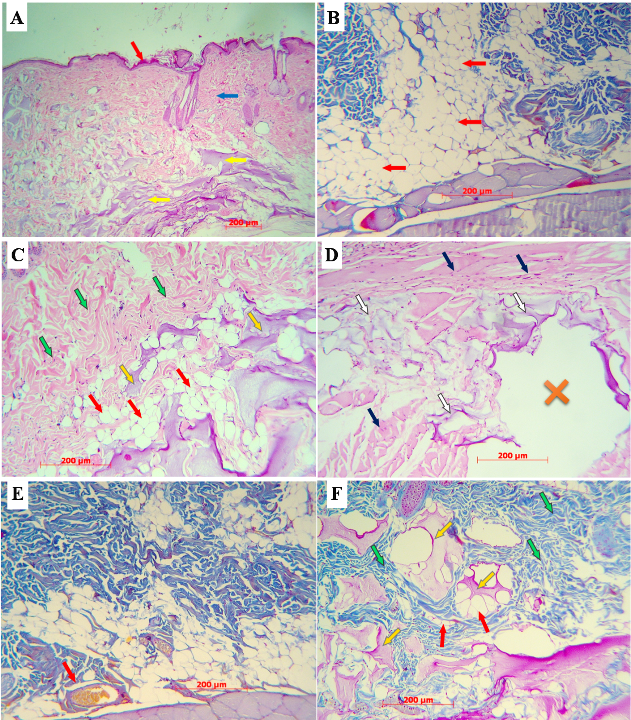
Figure 2. Histological sections. A – Skin of Group 4 animal on Day 30; the epidermis is shown by the red arrow, the dermis is pointed to by the blue arrow, the gel is indicated by the yellow arrows, stained with hematoxylin and eosin. B – dermis and hypodermis of Group 3 animal on Day 30; diffuse distribution of adipose tissue (red arrows), stained according to Mallory’s trichrome method. C – uneven distribution of the gel in Group 4 animal (Day 180), dermal fibers (green arrows), fat cells (red arrows), gel (filler) (yellow arrows); stained with hematoxylin and eosin. D – cavities in the dermis after filler implantation (Day 180): cavity (marked with ×), dermal fibers (blue arrows) and gel (white arrows); stained with hematoxylin and eosin. E – dermis and hypodermis in a Group 3 rat (after 90 days): diffuse distribution of adipose tissue (red arrows), microvessels (red arrow); Mallory’s trichrome stain. F – integration of gel (yellow arrows) with surrounding tissues in Group 4 (after 90 days): dermal fibers (green arrows), fat cells (red arrows), Mallory’s trichrome stain.
There was no difference in the thickness of the granular layer between two control groups. According to the Mann–Whitney test, after 30 (p=0.02) and 180 (p=0.012) days in Group 3, and after 30 (p=0.01) and 180 (p=0.004) days in Group 4, the granular layer was thicker than in the control groups (Figure 1B, Table 1). This parameter value was lower in Group 3 vs. Group 4 on Day 90 (p<0.001). In the fat graft group, the thickness of the granular layer on Day 90 decreased compared to Day 30, but increased again on Day 180 after surgery (p<0.001). In the hyaluronic acid filler group, the granular layer was significantly thinner at 30 days postoperatively than at 90 and 180 days (p<0.001).
The thickness of the cornified layer did not differ between the control groups. According to the Mann-Whitney test, the thickness of this layer, compared with the negative and positive control groups, was higher only in Group 3 at 30 days postoperatively (p=0.005). At 90 days, the cornified layer was thicker in the hyaluronic acid filler group than in the lipofilling group (p=0.003). In Group 3, the thickness of this layer significantly decreased at 90 and 180 days vs. Day 30 (p<0.001; Figure 1C, Table 1).
The thickness of the dermis did not differ between the control groups. According to the Mann-Whitney test, it was higher on day 30 in Group 3 (Figure 2B) and on day 180 in Group 4 (p<0.001; Figure 2C), as well as at 90 and 180 days in Group 3 (p=0.018), and at 30 (p=0.014) and 180 (p<0.001) days in Group 4 (Figure 1D, Table 2).
Table 2. Changes in the thickness of the dermis and sebaceous gland area size at the recipient site after fat graft implantation and hyaluronic acid filler injection, M±SE
| Group | Day# | ||
| 30 | 90 | 180 | |
| Dermis thickness, µm | |||
| 1 | 371±33 | ||
| 2 | 390±49 | ||
| 3 | 514±37 | 483±53 | 524±67 |
| 4 | 532±67 | 566±28 | 577±22 |
| Sebaceous gland area size, µm2 | |||
| 1 | 3,794±224 | ||
| 2 | 3,653±394 | ||
| 3 | 3,036±235 | 4,055±395 | 3,486±628 |
| 4 | 3,968±218 | 4,336±274 | 4,234±125 |
Sebaceous gland area size did not differ between the control groups. According to the Mann-Whitney test, this parameter value significantly decreased vs. to the control group only on Day 30 postoperatively (p<0.001). In Group 4, on the contrary, it increased after 90 and 180 days after the introduction of a hyaluronic acid filler compared to the control data (p=0.02). In Group 3, an increase in the value of this parameter was noted on Day 90 vs. Day 30 (p<0.001), but after 180 days, the sebaceous gland area size did not differ from that on Day 30. In Group 4, a stable increase in the sebaceous gland area size was noted after 90 and 180 days compared to Day 30 after the introduction of a hyaluronic acid-based product (p=0.002; Figure 1E, Table 2).
According to the Mann-Whitney test, fat graft thickness significantly decreased on Day 90 postoperatively compared to Day 30 (p=0.041; Figure 1D, 1E). Gel thickness in Group 4 was greater than in Group 3 on days 30 (p=0.038), 90 (p=0.007), and 180 (p<0.001; Figure 1D).
In Group 4, 90 days after hyaluronic filler implantation, integration of the gel with the surrounding tissue was observed (Figure 2F). The implanted gel often had a bubbly or layered consistency. After 180 days, the thickness of the implanted hyaluronic acid gel in this group varied, as the gel was often unevenly distributed, forming patches (Figure 2C). Cavities frequently appeared after gel implantation (Figure 2D). They were not the result of gel degradation, as they were visible even early after implantation. After 180 days of hyaluronic acid filler implantation, no noticeable reduction in the thickness of the injected filler was observed vs. earlier measurements (Figure 1F).
Discussion
The epidermis of control animals was thinner than in experimental animals, and higher mitotic activity was observed in the basal keratinocytes of experimental rats. The number of rows of granular keratinocytes and the size of keratohyalin granules in their cytoplasm are indicators of epidermal keratinization activity [7]. In our study, epidermal thickening in the experimental groups was due to the stimulation of tissue proliferation by growth factors from the SVF cells, which was confirmed by an increase in Ki-67 protein expression at the recipient site after fat grafting [8].
It should be noted that all animals in the control group lacked subcutaneous adipose tissue. In rats of Group 3, a subcutaneous fat base formed, which was likely caused by the activation of pericytes in the hypodermis and their differentiation into adipocytes [9]. Moreover, only some individuals in Group 4 exhibited foci of adipose tissue in the hypodermis. According to experimental data by some authors, hyaluronic acid fillers can stimulate the formation of adipose tissue [10], which is also confirmed by the present study. A hyaluronic acid filler was detected at all times after the experiment, but it was not always distributed evenly in the skin after 30 and 90 days, making it difficult to measure its thickness; this finding is confirmed by the data collected by other authors [11]. Furthermore, a hyaluronic acid filler can inhibit lipolysis [12].
Milled autologous fat and hyaluronic filler integrate well with surrounding skin tissues, thus promoting the formation of new microvessels. Our recent study demonstrated that autologous fat grafting stimulates the expression of VEGF A, which, in turn, promotes better fat engraftment and stimulation of neoangiogenesis and adipogenesis [13]. According to many authors, milled fat contains SVF [12], from which future endothelial cells (fibroblast-like precursors capable of accumulating lipids and acquiring the morphology of fat cells) differentiate [3, 14]. In the autologous fat transfer group in this study, the preservation of adipose tissue by Day 30 after the experiment can be explained by the release of inflammatory mediators that initiate both the activity of the SVF [14] and stress-related mechanisms. In contrast, hyaluronic acid is not foreign to the skin, as it is part of the extracellular matrix (glycosaminoglycans) and does not cause immune responses upon implantation [15, 16], although it sometimes stimulated the formation of adipose tissue foci in some animals of Group 4, which is supported by the data of some studies [10].
The preference for using a particular implant largely depends on the specified clinical goals. Implanted fat successfully takes root in the recipient area and can provide long-term favorable outcomes in soft tissue volume restoration. However, there is evidence of a deforming effect of fat grafts when they are used to correct protruding scars, which can lead to visible skin deformation [2]. Despite its effectiveness, lipofilling in visually accessible skin zones should be performed with caution [17], including due to possible local complications such as erythema, edema, and hematoma at the fat harvesting area or fat grafting site [18].
Hyaluronic gel implantation is characterized by a more predictable outcome [19], since hyaluronic acid (as a component of the extracellular matrix) is distributed evenly, without causing inflammatory responses or complications. Some authors pointed out that the duration of its clinical and aesthetic effects is much shorter than that of transplanted fat grafts [20].
Conclusion
The need to evaluate morphological changes in surrounding tissues at the recipient site during lipofilling simulations and the use of hyaluronic acid fillers is pathogenetically substantiated in this article. For instance, in an experiment using autologous fat transfer, we observed a more uniform distribution of adipose tissue and a reduction in the area size of sebaceous glands, compared with hyaluronic acid fillers. The latter contribute to an increase in the thickness of the epidermis (including due to the growth of the granular layer), the thickness of the dermis, and the filler per se in the subcutaneous space, compared to lipofilling.
It should be noted that the choice of a specific type of implant is determined primarily by the specific clinical needs, while it is recommended to consider the detected changes in the surrounding tissues and the mechanisms that caused them as well.
Conflict of interest
The authors declare no conflicts of interest.
Author contributions
I.V. Kastyro, A.G. Korolev, and A.A. Tsymbal designed the study; E.A. Lavrentyeva and A.G. Korolev conducted the experiment; A.G. Korolev and E.A. Lavrentyeva performed statistical analysis; E.A. Lavrentyeva, A.G. Korolev, and I.V. Kastyro wrote the article; I.B. Ganshin, M.V. Barannik, P.V. Sarygin, and A.A. Tsymbal edited the text.
Funding
The authors declare no external funding for the study or publication of this article.
Bioethics compliance
The study was conducted in accordance with the ethical standards for the treatment of animals adopted by the European Convention for the Protection of Vertebrate Animals used for Experimental and other Scientific Purposes.
Quartey A, Ezekwe U, Callender V. Hyaluronic acid delayed filler complications: A racial group analysis and call to action. Arch Dermatol Res 2025; 317(1): 690. https://doi.org/10.1007/s00403-025-04125-x.
Nalcı Baytaroğlu H, Hoşal MB. Complications of periorbital cosmetic hyaluronic acid filler injections: A major review. Turk J Ophthalmol 2025; 55(5): 276-286. https://doi.org/10.4274/tjo.galenos.2025.45213.
Bora P, Majumdar AS. Adipose tissue-derived stromal vascular fraction in regenerative medicine: A brief review on biology and translation. Stem Cell Res Ther 2017; 8(1): 145. https://doi.org/10.1186/s13287-017-0598-y.
Zheng Z, Lei X, Yang Y, et al. Changes in human fat injected alongside hyaluronic acid in the backs of nude mice. Aesthet Surg J 2021; 41(6): NP631-NP642. https://doi.org/10.1093/asj/sjaa351.
Qu Z, Koga H, Tsuji K, et al. Hyaluronic acid sheet transplantation attenuates infrapatellar fat pad fibrosis and pain in a rat arthritis model. J Orthop Res 2023; 41(11): 2442-2454. https://doi.org/10.1002/jor.25580.
Fanian F, Deutsch JJ, Bousquet MT, et al. A hyaluronic acid-based micro-filler improves superficial wrinkles and skin quality: A randomized prospective controlled multicenter study. J Dermatolog Treat 2023; 34(1): 2216323. https://doi.org/10.1080/09546634.2023.2216323.
Hoober JK, Eggink LL. The discovery and function of filaggrin. Int J Mol Sci 2022; 23(3): 1455. https://doi.org/10.3390/ijms23031455.
Moroz SE, Lavrentieva EA, Khlystalov MV, et al. Pathomorphological changes in tissues with various methods of transplantation of adipose tissue (experimental study). Head and Neck. Russian Journal. 2024; 12(4): 41-49. (In Russ.) https://doi.org/10.25792/HN.2024.12.4.41-49.
Shi Z, Xiong S, Hu R, et al. The Notch-PDGFRβ axis suppresses brown adipocyte progenitor differentiation in early post-natal mice. Dev Cell 2024; 59(10): 1233-1251.e5. https://doi.org/10.1016/j.devcel.2024.03.012.
Crowley JS, Kream E, Fabi S, Cohen SR. Facial rejuvenation with fat grafting and fillers. Aesthet Surg J 2021; 41(Suppl. 1): S31-S38. https://doi.org/10.1093/asj/sjab014.
Nadra K, André M, Marchaud E, et al. A hyaluronic acid-based filler reduces lipolysis in human mature adipocytes and maintains adherence and lipid accumulation of long-term differentiated human preadipocytes. J Cosmet Dermatol 2021; 20(5): 1474-1482. https://doi.org/10.1111/jocd.13794.
Ehlinger-David A, Gorj M, Braccini F, et al. A prospective multicenter clinical trial evaluating the efficacy and safety of a hyaluronic acid-based filler with Tri-Hyal technology in the treatment of lips and the perioral area. J Cosmet Dermatol 2023; 22(2): 464-472. https://doi.org/10.1111/jocd.15169.
Khlystalov MV, Kastyro IV, Popadyuk VI, Ganshin IB. Diameter of blood capillaries in biological objects after fat grafting in the long term. Head and Neck. Russian Journal. 2026; 14(1): 91-97. (In Russ.) https://doi.org/10.25792/HN.2026.14.1.91-97.
Gandolfi S, Pileyre B, Drouot L, et al. Stromal vascular fraction in the treatment of myositis. Cell Death Discov 2023; 9(1): 346. https://doi.org/10.1038/s41420-023-01605-9.
Marinho A, Nunes C, Reis S. Hyaluronic acid: A key ingredient in the therapy of inflammation. Biomolecules 2021; 11(10): 1518. https://doi.org/10.3390/biom11101518.
Wu GT, Kam J, Bloom JD. Hyaluronic acid basics and rheology. Clin Plast Surg 2023; 50(3): 391-398. https://doi.org/10.1016/j.cps.2022.12.004.
Klingenstein A, Garip-Kuebler A, Muth DR, Hintschich C. A prospective randomized pilot study evaluating the scar outcome after gluteal dermis fat graft with and without kinesiotaping. Int Ophthalmol 2022; 42(8): 2563-2571. https://doi.org/10.1007/s10792-022-02304-7.
Le JM, Bosworth JW, Honeywell B, et al. Adipose grafting for volume and scar release. Ann Plast Surg 2021; 86(6S Suppl. 5): S487-S490. https://doi.org/10.1097/SAP.0000000000002873.
Salwowska NM, Bebenek KA, Żądło DA, Wcisło-Dziadecka DL. Physiochemical properties and application of hyaluronic acid: A systematic review. J Cosmet Dermatol 2016; 15(4): 520-526. https://doi.org/10.1111/jocd.12237.
Tabassum N, Chowdary Jasthi V, Al Salem A, Kumar SM. Perspectives and challenges in lip rejuvenation: A systematic review. Eur Rev Med Pharmacol Sci 2023; 27(19): 9043-9049. https://doi.org/10.26355/eurrev_202310_33929.