Materials and Methods: TREC and KREC values were dynamically evaluated in 203 children born at 22–41 weeks of gestation. Dried blood spots applied to Guthrie cards served as the background TREC and KREC levels, while venous blood was used for repeat determinations and 79 children whose values were outside the reference ranges underwent repeat assessment of the markers at two and four weeks from the initial sampling date.
Results: In immunocompromised newborns, the values of the target markers gradually normalize with each week of postnatal development. TREC values are statistically significantly affected by maternal history factors such as congenital anomalies of the kidneys (p=0.008), spontaneous abortions (p=0.004), chronic vaginitis (p=0.015), nicotine addiction (p<0.001), and alcohol abuse (p=0.029), while KREC values are significantly affected by chronic pyelonephritis (p=0.028) and bacterial vaginosis (p=0.02).
Conclusion: With an increase in the volume of immunocompetent cells, there is a commensurate increase in the number of TRECs and KRECs, as a result of which children with borderline values of the target markers are classified as conditionally healthy.
Introduction
The immune system is a complex of organs and tissues regulating the functioning of the body’s vital systems. If, due various congenital defects, the immune system is unable to withstand external aggressive impact leading to a generalized infectious process, then the likelihood of severe complications is high [1]. This is especially true in premature newborns, because sepsis is the leading cause of neonatal mortality in them [2].
The general gestational immaturity of the major organs and systems complicates the differential diagnosis of primary immunodeficiency conditions. This circumstance, in conjunction with high neonatal mortality rates, has prompted the contemporary scientific community to conduct comprehensive studies of the immune system in premature neonates [3, 4]. For instance, since the beginning of 2023, expanded neonatal screening has been introduced in the Russian Federation to identify severe inborn errors of immunity (IEI) [4]. This method involves determining the number of the following molecules: T-cell receptor excision circles (TRECs) formed in the course of the maturation of T lymphocytes and kappa-deleting recombination excision circles (KRECs) appearing during the development of B lymphocytes. TRECs reflect the activity of T cells recently released from the thymus, while KRECs reflect the activity of B cells recently released from the bone marrow [5]. This method is simple to perform, and it exhibits high sensitivity and specificity [6].
Although the described analysis is very important for diagnosing primary immunodeficiency (when TREC and KREC values fall below 100 copies per 105 leukocytes per µL), it can also identify children whose values are within the conditionally normal range (100-450 copies for TREC and 100-250 copies for KREC per 105 leukocytes per µL, respectively). Such newborns fall into the category of conditionally healthy since they do not require urgent in-depth immunological examination. However, it is also impossible to fully exclude various types of IEI in them until the values of the assessed markers are back to normal [3].
Objective: Dynamic assessment of TREC and KREC values in immunocompromised neonates, along with identification of noteworthy anamnestic factors that may influence the values of these markers.
Materials and Methods
We conducted a prospective single-center continuous study that included 203 newborns with a gestational age of 22-41 weeks. They were delivered at the Perinatal Center of the Voronezh Oblast Clinical Hospital No. 1 from 2024 through 2025 (the total sample size was not precalculated). Assignment of neonates to study groups was based on the gestational age at the time of birth: Group 1 with children born at 22-27 weeks of gestation (n=39), Group 2 with children born at 28-30 weeks (n=57), Group 3 with children born at 31-33 weeks (n=48), Group 4 with children born at 34-36 weeks (n=34), and Group 5 with children born at 37-41 weeks (n=25). The inclusion criteria were as follows: 1) Children born at 22-41 weeks; 2) The presence of informed voluntary consent for data processing and their use for scientific purposes signed by the patient’s legal representative. The exclusion criteria for the study were: 1) Inadequate blood sample; 2) The presence of congenital defects in the child; 3) The presence of transfusions of various blood components the day before collection of the blood sample for the analysis.
The study was conducted in two stages. In Stage 1, TREC and KREC values were determined from dried capillary blood spots applied to Guthrie cards. Blood was collected as part of the extended neonatal screening program in compliance with the Order No. 274n of the Russian Federation Ministry of Healthcare of April 21, 2022, “On Approval of the Procedure for Providing Medical Care to Patients with Congenital and (or) Hereditary Diseases.” Blood samples were collected from full-term neonates 24-48 h after birth and from premature newborns 144-168 h after birth. DNA was extracted from the obtained biomaterial (dried blood spots were used during Stage 1 of the study, while venous blood was used during Stage 2). The Extra-DNA-Bio reagent kit (AlkorBio Company LLC, Russia) was employed according to the manufacturer’s instructions. TREC and KREC were determined by real-time polymerase chain reaction using the IMMUNO-BIT reagent kit (ABT-Test LLC, Russia). Primers specific for the human ABL gene region served as a control. TREC and KREC were determined strictly according to the manufacturer’s guidelines. The number of TREC (KREC) copies was calculated using the following formula:
TREC/KREC copy number = [TREC (KREC) copy number per mL] / (ALB copy number) × 200,000.
TREC and KREC values (the number of copies per 105 leukocytes per µL) are usually interpreted as follows:
TREC: 1) normal values (>450), 2) immunocompromised newborns (100-450 copies), 3) primary immunodeficiency/IEI (<100 copies);
KREC: 1) normal values (>250 copies), 2) immunocompromised newborns (100-250 copies), 3) primary immunodeficiencies/ IEI (<100 copies) [3].
To identify factors that statistically significantly affect TREC and KREC values, we performed a retrospective assessment of anamnestic data collected from medical documentation (anamneses of newborn development) of newborns with gestational age of 22–36 weeks (n=178). Also, we analyzed maternal anamnesis data, including adverse somatic and obstetric histories, and complicated pregnancy.
During Stage II of the study, infants (n=73) whose TREC and KREC levels did not reach the reference range (100 to 450 copies for TREC/100 to 250 copies for KREC per 105 leukocytes per μL) underwent a repeat assessment of the marker levels two and four weeks after the initial blood collection date. Venous blood collected from an intact vein on an empty stomach served as the material for repeat TREC and KREC level assessment.
The study was conducted in accordance with Good Clinical Practice standards and the principles of the Declaration of Helsinki. All stages of the study were approved by the Ethics Committee of the N.N. Burdenko Voronezh State Medical University of the Russian Federation Ministry of Healthcare (Protocol No. 4 of October 16, 2024). For each newborn enrolled in our study, we obtained the informed consent from the parents.
Statistical data processing was performed using StatTech v.4.8.5 (StatTech LLC, Russia). Quantitative indicators were assessed for compliance with normal distribution using the Shapiro–Wilk test (for fewer than 50 subjects) or the Kolmogorov–Smirnov test (for 50 subjects or more). In the absence of normal distribution, quantitative data were described using the median and the lower and upper quartiles (Me [Q1-Q3]). Two groups were compared with each other by a quantitative parameter using the Mann–Whitney U test (for non-normal distributions), three or more groups were compared using the Kruskal–Wallis test, and post-hoc comparisons were performed using the Dunn’s test with Holm’s correction. Percentage comparisons in multivariate contingency tables were carried out with the Pearson’s χ2 test, while Pearson’s χ2 test with Holm’s correction was employed for post-hoc comparisons. When comparing three or more dependent populations with distributions other than normal, we used the nonparametric Friedman test with post-hoc comparisons via the Conover-Iman test with Holm’s correction. The statistical significance of parameter changes over time was assessed using the Fisher’s F test. Differences were considered statistically significant at p<0.05.
Results
TREC and KREC values in newborns of various gestational ages are presented in Table 1.
Table 1. Counts of T-cell receptor excision circles (TRECs) and kappa-deleting recombination excision circles (KRECs) in the general cohort of newborns, copies per 105 leukocytes per µL
| Indicator value | Study groups, weeks | p | |||||
| 22-27th, n=39 | 28-30th, n=57 | 31-33rd, n=48 | 34-36th, n=34 | 37-41st, n=25 | |||
| TREC | Me [Q1-Q3] | 520.0 [222.5-742.0] | 910.0 [376.0-1,201.0] | 635.0 [305.5-1,311.0] | 807.0 [232.5-1,224.0] | 675.0 [334.0-1,475.0] | 0.024 |
| KREC | 597.0 [294.0-782.0] | 878.0 [609.0-1,430.0] | 876.0 [302.0-1,686.0] | 935.0 [637.25-1,777.25] | 991.0 [480.0-2,191.0 | 0.005 | |
We revealed that the lowest TREC and KREC values were observed in neonates born at 22-27 weeks of gestation. With increasing gestational age, the values of the target markers gradually increased, reaching their maximum at 36 weeks for TREC and at 41 weeks for KREC. The most pronounced increase (on average, 1.5-fold) in the values of the assessed markers was observed at 28-30 weeks of gestation. It is also noteworthy that the dynamics of KREC values was more linear.
Besides that, we noted that in each study group, approximately one-third of all infants exhibited values of the assessed markers that were beyond the reference range (the lower the gestational age, the higher the percentage of newborns with atypical values). The distribution of infants with such deviating TREC and KREC values across the study groups is demonstrated in Table 2.
Table 2. Distribution of newborns with abnormal values of T-cell receptor excision circles (TRECs) and kappa-deleting recombination excision circles (KRECs), copies per 105 leukocytes per µL
| Indicator | Category | Study groups, weeks | р | ||||
| 22-27th, n=39 | 28-30th, n=57 | 31-33rd, n=48 | 34-36th, n=34 | 37-41st, n=25 | |||
| Number of newborns, count (%) | |||||||
| TREC | <100 | 5 (12.8) | 1 (1.8) | 2 (4.2) | 0 (0.0) | 0 (0.0) | 0.151 |
| 100-450 | 12 (30.8) | 17 (29.8) | 15 (31.2) | 13 (38.2) | 9 (36.0) | ||
| >450 | 22 (56.4) | 39 (68.4) | 31 (64.6) | 21 (61.8) | 16 (64.0) | ||
| KREC | <100 | 4 (10.3) | 2 (3.5) | 3 (6.3) | 2 (5.9) | 1 (4.0) | 0.882 |
| 100-450 | 3 (7.7) | 6 (10.5) | 6 (12.5) | 3 (8.8) | 2 (8.0) | ||
| >450 | 32 (82.0) | 49 (86.0) | 39 (81.2) | 29 (85.3) | 22 (88.0) | ||
Due to the fact that many children were in the immunocompromised neonatal group (isolated decreases in KREC were observed in 13 children, while isolated decreases in TREC were noted in 60 children, and simultaneous decreases in TREC and KREC were characteristic of 6 children), a repeat assessment of the markers was performed in 79 neonates.
We obtained the following results. In immunocompromised children, the median TREC values were 277.50 [195.0-340.75] at the initial blood collection, 370.00 [298.25-459.0] after two weeks, and 459.00 [349.0-534.75] after four weeks. The median KREC values were 166.0 [135.0-197.0] initially, 212.0 [188.0-247.0] after two weeks, and 291.00 [255.0-311.0] after four weeks. The differences were statistically significant (p<0.001 for both TREC and KREC). Taking these data into account, we conclude that with increasing postnatal age, a gradual normalization of the assessed parameters occurs. A graphical interpretation of the obtained results is presented in Figures 1 and 2.

Figure 1. Dynamics of counts of T-cell receptor recombination excision circles in immunocompromised children.
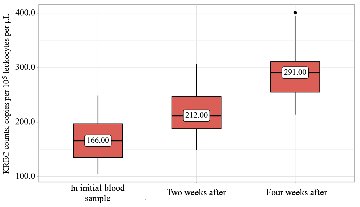
Figure 2. Dynamics of counts of kappa-deleting recombination excision circles in immunocompromised children.
In Figures 1 and 2, the box plot represents the interquartile range (the lower limit of the box represents the 25th percentile, the upper limit of the box represents the 75th percentile). The upper whisker represents the maximum value in the subgroup that is not an outlier (estimated by 95% of the included data). The lower whisker represents the minimum value in the subgroup that is not an outlier (estimated by 95% of the included data). The line in the middle of the box represents the median. The dot represents an outlier.
To reveal the main factors that can lead to reduced levels of the assessed markers, we examined the maternal anamneses of the examined newborns. We discovered that the presence of such pathologies in the mother as congenital anomalies of the kidneys (p=0.008), spontaneous abortions (p=0.004), chronic vaginitis (p=0.015), nicotine addiction (p<0.001), and alcohol abuse (p=0.029) statistically significantly influence the TREC values, while chronic pyelonephritis (p=0.028) and bacterial vaginosis (p=0.020) impact the KREC values. Due to the presence of statistically significant relationships, these pathological conditions can be considered as risk factors leading to a decrease in the values of the assessed markers. A detailed description of the anamnestic factors is presented in Table 3.
Table 3. Anamnestic factors significantly influencing counts of T-cell receptor excision circles (TRECs) and kappa-deleting recombination excision circles (KRECs), copies per 105 leukocytes per µL
| Indicator | Category | Absence (–) / presence (+) | Me [Q1-Q3] | n | p |
| TREC | Congenital anomalies of the kidneys | – | 659.0 [330.5-1177.0] | 172 | 0.008 |
| + | 176.0 [155.0-242.5] | 6 | |||
| Nicotine addiction | – | 795.0 [388.5-1311.5] | 108 | <0.001 | |
| + | 494.5 [218.5-910.0] | 70 | |||
| Alcohol abuse | – | 659.0 [311.0-1180.0] | 169 | 0.029 | |
| + | 273.0 [205.0-589.0] | 9 | |||
| Spontaneous abortion | – | 785.5 [376.0-1197.5] | 132 | 0.004 | |
| + | 413.0 [228.5-756.5] | 46 | |||
| Chronic vaginitis | – | 661.5 [308.5-1177.0] | 172 | 0.015 | |
| + | 281.0 [110.5-450.5] | 6 | |||
| KREC | Chronic pyelonephritis | – | 854.0 [418.5-1447.5] | 146 | 0.028 |
| + | 539.0 [302.0-910.5] | 32 | |||
| Bacterial vaginosis | – | 878.0 [454.0-1417.0] | 127 | 0.020 | |
| + | 662.0 [198.5-1145.5] | 51 |
Discussion
The pattern we revealed, associated with an increase in TREC and KREC values commensurate with the gestational age, can be also seen in data of some recent publications. For example, a study that included 177,277 newborns noted that with increasing gestational age, the concentrations of the assessed markers increase [7]. Similar data were obtained by M. Giżewska et al., who demonstrated that TREC and KREC values are directly correlated with gestational age. It is also interesting that in that study, the increase in the marker values did not occur linearly, but rather with the development of various peaks (the most significant of which developed between the 28th and 30th weeks) [5]. This pattern is explained by the physiological growth of the child’s body (with an increase in the gestational age, there is a gradual development of the central and peripheral organs of the immune system, which is manifested by an increase in the number of the main immunocompetent cells) [4].
Our study also demonstrated that TREC levels are statistically significantly affected by maternal anamnestic factors such as congenital anomalies of the kidneys, spontaneous abortions, chronic vaginitis, nicotine addiction, and alcohol abuse; while KREC levels are affected by chronic pyelonephritis and bacterial vaginosis. This is consistent with current published data. E.g., a number of studies have found that the incidence of severe intrauterine infection was higher in newborns of mothers whose pregnancy was complicated by any infectious process vs. children of apparently healthy women (severe intrauterine infection leads to a decrease in TREC and KREC levels) [8]. D.A. Kashatnikova et al. noted in their study that the values of the assessed markers were several times lower in patients with severe pneumonia compared to patients without this pathology. It is also noteworthy that the authors considered a critical reduction in TREC and KREC levels a marker of a high risk of subsequent development of a severe septic process [9]. M. Von-Chamier et al. found that long-term nicotine exposure negatively affects the body of pregnant animals (high doses of nicotine potentiate the progression of intrauterine infection, which is also manifested by a reduction in TREC and KREC levels) [10]. T. Deak et al. confirmed that alcohol has a significant negative impact on the neuropsychic development of the unborn child and also leads to the formation of pronounced immune dysfunction [11].
We also discovered that in immunocompromised populations, there is a gradual normalization of T and B cell immunity (in particular, TREC and KREC) with increasing postnatal age. The available data in recent publications are very scarce and contradictory. For example, a number of authors have shown that most premature infants, who represent one of the most significant immunocompromised population groups, do not exhibit any significant health abnormalities by the end of their first year of life (not a single confirmed case of acute infectious disease was recorded in this category of children after discharge from hospital during their first year of life) [12]. In contrast, E.I. Penkina et al. confirmed that an initial decrease in TREC and KREC levels can be a specific predictor of the development of various pathologies, in particular sudden infant death syndrome and upper respiratory tract diseases (the latter is manifested by up to 12 episodes of acute respiratory infections annually) [13].
Conclusion
The conducted study expands our knowledge of the differential diagnosis of conditions not associated with the development of severe immunodeficiency, but characterized by a reduction in the content of the main markers of T and B cell immunity. Further development of this scientific direction will improve the results of nursing newborns, especially for premature babies.
Conflict of interest
The authors declare no conflicts of interest related to the publication of this article.
Author contributions
All authors contributed equally to the preparation of this publication.
Informed consent for publication
The authors obtained written consent from the patients’ legal representatives for the analysis and publication of their medical data.
Funding
The authors received no external funding for the study and publication of this article.
Compliance with ethical principles
The study protocol was approved by the local ethics committee. The approval and procedure for conducting the research fully complied with the principles of the Declaration of Helsinki.
Olin AJ, Henckel EA, Chen Y, et al. Stereotypic immune system development in newborn children. Cell 2018; 174(5): 1277-1292.e14. https://doi.org/10.1016/j.cell.2018.06.045.
Kariniotaki CA, Thomou CD, Gkentzi DI, et al. Neonatal sepsis: A comprehensive review. Antibiotics (Basel) 2024; 14(1): 6. https://doi.org/10.3390/antibiotics14010006.
Prodeus AP, Odinayeva ND, Voronin SV, et al. Guidelines for routing, monitoring and management tactics of immunocompromised children at risk and patients with congenital immune defects. Pediatria n.a. G.N. Speransky 2024; 103(6): 218-245. (In Russ.) https://doi.org/10.24110/0031-403X-2024-103-6-218-245.
Cheremokhin DA, Shinwari H, Deryabina SS, et al. Analysis of the TREC and KREC levels in the dried blood spots of healthy newborns with different gestational ages and weights. Acta Naturae 2022; 14(1): 101-108. (In Russ.) https://doi.org/10.32607/actanaturae.11501.
Giżewska MJ, Durda K, Winter TA, et al. Newborn screening for SCID and other severe primary immunodeficiency in the Polish-German transborder area: Experience from the first 14 months of collaboration. Front Immunol 2020; 11(1): 1948. https://doi.org/10.3389/fimmu.2020.01948.
Marinova MА, Georgyeva AR, Yordanova VG, et al. Implementation of TREC/KREC detection protocol for newborn SCID screening in Bulgaria: A pilot study. Cent Eur J Immunol 2022; 47(4): 339-349. https://doi.org/10.5114/ceji.2022.124396.
Rechavi EА, Lev AЕ, Simon AJ, et al. First year of Israeli newborn screening for severe combined immunodeficiency-clinical achievements and insights. Front Immunol 2017; 8(1): 1448. https://doi.org/10.3389/fimmu.2017.01448.
Shadeeva YuA, Gurjeva VA. Risk factors for intrauterine infection in extremely preterm and very preterm birth, complicated by the rupture of amniotic membranes. Mother and Baby in Kuzbass 2019; (4): 33-39. (In Russ.) https://www.elibrary.ru/arqnrq.
Kashatnikova DA, Khadzhieva MB, Kolobkov DS, et al. Pneumonia and related conditions in critically ill patients-insights from basic and experimental studies. Int J Mol Sci 2022; 23(17): 9896. https://doi.org/10.3390/ijms23179896.
Von-Chamier MА, Reyes LА, Hayward LF, et al. Nicotine induces maternal and fetal inflammatory responses which predispose intrauterine infection risk in a rat model. Nicotine Tob Res 2021; 23(10): 1763-1770. https://doi.org/10.1093/ntr/ntab080.
Deak TА, Kelliher KT, Wojcik HJ, Gano A. Prenatal and adolescent alcohol exposure programs immunity across the lifespan: CNS-mediated regulation. Pharmacol Biochem Behav 2022; 216: 173390. https://doi.org/10.1016/j.pbb.2022.173390.
Shilova NA, Kharlamova NV, Matveeva EA. Dynamics of health status and outcomes of perinatal pathology in deeply premature babies at the age of three. Vestnik Ivanovskoi Meditsinskoi Akademii 2023; 28(3): 23-27. (In Russ.) https://doi.org/10.52246/1606-8157_2023_28_3_23.
Penkina NI, Ermolaeva NI, Kudlay DA, Prodeus AP. Retrospective diagnosis of immunoregulatory disorders in children requiring dispensary monitoring. Vrach (The Doctor) 2023; 34(5): 88-93. (In Russ.) 2023; 34(5): 88-93. https://doi.org/10.29296/25877305-2023-05-16.