Materials and Methods. A morphological study was performed on 60 laboratory sexually mature rats. The study materials were implant specimens from four groups with different types of VT6 titanium alloy coatings.
Results. Biological compatibility of all implant specimens was confirmed. In the uncoated implant group, no significant bone formation was observed, and implant stability remained low by Month 2 (2.9±1.1 pts on the implant stability scale). Chitosan-coated implants exhibited low stability (1.8±0.1 pts by Week 2), but significant stimulation of osteogenesis was observed. In the antibacterial coating group, good integration and minimal implant mobility were noted (3.4±0.3 pts after one), and the antibacterial coating was preserved for two weeks.
Conclusion: Hydroxyapatite and chitosan coatings contributed to quicker osseointegration via stimulating angiogenesis, stromal collagen synthesis, and bone formation, while maintaining full biocompatibility and rapid biodegradation. The antibacterial coating ensured rapid relief of inflammation in the postoperative wound area.
Introduction
In contemporary reconstructive surgery, there is a dire need for implants covered with biologically active coatings. Their role can vary depending on the implant’s application, lifespan, function, and purpose. Such combined bioconstructs are specifically in demand in skeletal surgery – viz., in traumatology and orthopedics, maxillofacial surgery, and dental implantology [1–3].
The main requirements for such implants are their biocompatibility, absence of cytotoxicity, and no immune responses [1, 3]. For musculoskeletal surgery, reliable integration of implants with the host tissue is essential. For devices intended for bone fragment fixation, bone tissue becomes such a tissue of the receiving bed when external implants are stabilized. A number of coatings can enhance the osseointegration of implanted metal structures [4, 5].
Among materials providing this function, we should mention calcium hydroxyapatite (HAp). In recent years, numerous modifications of this coating have been developed, along with its manufacturing methods, application to the implant, and fixation. Experimental and clinical studies have demonstrated its safety and effectiveness for the integration of implants with bone tissue [6–8].
However, a number of issues related to the surface treatment with HAp, the degree of porosity of such coatings, and the possibility of combining the coating with other biologically active substances remain unresolved [9]. These issues have been consistently addressed in recent studies. In particular, given the relatively high infection rate (up to 10%) during revision arthroplasty of large joints, instilling such implant coatings with antibacterial agents is a pressing issue [10]. Controlling the gradual release of medicinal substances, which ensures the prolonged action of such coatings, is a challenge.
Modifications of HAp containing antibiotics used in reconstructive surgery are known [11]. At the same time, many studies present problems associated with the rapid release of antibacterial substances, absence of their prolonged effect, the complexity of the interaction of drugs and HAp during their combination and processing, and even the deterioration of the osseointegration properties of HAp when attempting to combine the coating with other medicinal drugs [12].
Hence, monitoring the development of combined coatings, assessing their initial osseointegration properties and biocompatibility, as well as monitoring changes in the quality of these effects when other coatings and drugs are employed, is of interest for the development of new metal structures in reconstructive surgery.
The goal of our study was to evaluate the safety and osseointegration properties of experimental implant specimens with different types of coatings in an in vivo experiment in rats.
Materials and Methods
This study was conducted at the vivarium of the BioTech Research Institute at Samara State Medical University, Ministry of Healthcare of the Russian Federation.
A scapula defect was simulated in animals. Our in vivo experiment assessed the safety and osseointegration properties of implants with different coating types via macroscopic and morphological examination.
The study materials were VT6 titanium implants manufactured using selective laser sintering technology (Pavlov First St. Petersburg State Medical University, Ministry of Healthcare of the Russian Federation). The implant specimens were distributed among four groups:
I – VT6 titanium implants without coatings;
II – implants with a HAp coating (National Research Tomsk Polytechnic University);
III – implants with a HAp coating additionally coated with a newly developed antibacterial agent (Skolkovo Institute of Science and Technology);
IV – implants with a coating containing chitosan (Department of Polymer and Crystal Physics, Lomonosov Moscow State University).
All specimens were sterilized by standard procedures used in medical clinics, including the use of ɣ-radiation.
The morphological study was performed on 60 mature laboratory Wistar rats, whose weight at the beginning of the experiment was, on average 270 g (range: 260–280 g).
For surgical interventions on animals and their care in the vivarium, we complied with the European Convention for the Protection of Vertebrate Animals used for Experimental and other Scientific Purposes (ETS No. 123, Strasbourg, March 18, 1986), the Interstate Standard (GOST 33044-2014) Principles of Good Laboratory Practice, the Russian Federal Standard GOST 33216-2014 Guidelines for the Care and Maintenance of Laboratory Animals. Rules for the Care and Maintenance of Laboratory Rodents and Rabbits, and the Sanitary and Epidemiological Requirements for the Design, Equipment, and Maintenance of Experimental Biological Clinics (Vivariums) (SP 2.2.1.3218-14).
The criteria for selecting animals for the experiment were the same age of individuals (6–7 months) and the absence of diseases. Rats were kept and maintained in a vivarium with a balanced light, water, and food regimens in accordance with Good Laboratory Practice.
All surgical procedures were performed under intramuscular anesthesia using a mixture of Zoletil 100 (Virbac SA, France) at a dosage of 0.03 mg and Rometar (Bioveta, Czech Republic) at a dosage of 0.08 mg. The animal was then secured on the operating table. Sterile surgical instruments were used for the experiment. All procedures were performed under aseptic conditions using antiseptic agents. To study the reactions of surrounding tissues to the tested implants, we employed their intraosseous implantation near the spine of the scapula.
Animals were withdrawn from the experiment by administering an overdose of Zoletil intramuscularly. Disarticulation of the scapulae was performed on the operating table. Animal bone specimens were subjected to 12% formalin fixation for at least three days and then rinsed under running water for 18 hours. The specimens were decalcified using conventional methods.
Macroscopic assessment of the implantation zone. During the macroscopic examination, the condition of the peri-implant tissues was assessed. The presence or absence of inflammation in the postoperative wound area and, later, the scar, as well as soft tissue changes, were noted: the presence of adhesions, excessive scar tissue growth around the implant, migration, implant mobility, or, conversely, a pronounced osseointegration around it.
To objectively interpret the results of evaluating macro specimens over time, we developed the scoring criteria described below.
To evaluate the macro specimen in terms of implant mobility in the implant bed (bone), the following scoring system was employed:
• No visible implant mobility in the implant bed (bone) (4 pts);
• Implant mobility in one horizontal plane (3 pts);
• Implant mobility in two horizontal planes (2 pts);
• Implant mobility in the vertical plane (1 pt).
The following scoring criteria were used to assess the macrostructure of the newly formed tissue complex in the implant area:
• Bone regenerate (bone callus) completely covers the end of the implant at the level of the host bone surface (4 pts);
• Bone regenerate partially covers the end of the implant, forming a bone cap (3 pts);
• The end of the implant is covered by a connective tissue capsule (2 pts);
• The end of the implant is exposed, and bone resorption is observed in the cervical portion of the implant (1 pt).
Morphological studies. For microscopic assessment of osseointegration, a bone specimen was collected. After fixation and decalcification of the bone tissue, the specimens were passed through increasing alcohol concentrations (70º alcohol for at least 12 hours, 96º alcohol for 4 to 18 hours, and 100º alcohol for 3 to 4 hours) and embedded in celloidin-paraffin blocks. Serial sections (5–10 µm thick,) were cut through the entire depth of the block using a Sakura Accu-Cut SRM 200 rotary microtome (Sakura Finetek, Japan).
For morphological evaluation, histological specimens were stained using conventional methods: hematoxylin and eosin, Van Gieson’s stain (picrofuchsin), and cresyl violet. To obtain reliable data and analyze surrounding tissue, every third section was stained. The obtained slides were analyzed and photographed using a Videotest instrumentation system with Morphology 5.2 software (Videotest LLC, St. Petersburg, Russia). A total of 546 slides were prepared.
Data were statistically processed using Microsoft Excel and BioStat spreadsheets: arithmetic means and their standard deviations were calculated. The normality of the distribution was assessed for all variables using the Shapiro-Wilk test. Results were processed using the Kruskal-Wallis and Newman-Keuls nonparametric tests for multiple samples. Differences were considered statistically significant at p<0.05.
Results
Results of a macroscopic examination of experimental implant specimens. The specimens were implanted into the scapular spine of rats, creating a hole with a burr to ensure the implant was firmly seated in the bone tissue. The wounds were sutured layer by layer. Consequently, implant contacted both the bone tissue and adjacent soft tissues, such as muscle and fascia. Animals were sacrificed at the end of Week 1, Week 2, Month 1, and Month 2 after implantation.
We assessed the integration of the implants with the implant bed (bone) and adjacent soft tissues, as well as the presence or absence of inflammation.
In no case did we observe postoperative wound suppuration. By the evening of the surgery day, the animals were already active, drinking and eating. The wounds were treated with antiseptic solutions and were not intentionally covered with dressings. In all comparison groups, the wounds closed via primary intention, forming postoperative scars. The local inflammatory response was present only for three – four days and was accompanied by slight hyperemia of the wound edges and moderate soft tissue swelling. After five days, these symptoms disappeared in all animals and did not recur.
Thermometry revealed a significantly early resolution of the local increase in skin temperature. We did not detect any complications in the early postoperative period in the comparison group rats.
Implants with a single calcium hydroxyapatite coating. Upon removal of the implant with a single HAp coating, we observed no inflammatory response in the peri-implant tissue at any time. The muscle tissue was pink and shiny. The fascia was visually unaffected, no scar tissue growth near the implant occurred, and no coating particles were detected in the tissue surrounding the implant.
The implant exhibited slight mobility when its end was clamped with tweezers by Week 2 of observation (2.2±1.1 pts on the implant stability scale; p>0.05); but by the end of Month 1, it was tightly fused with the adjacent bone and could not be displaced by the instrument (3.8±0.8 pts on the implant stability scale; p=0.032). Visual macroscopic assessment of bone formation revealed that by Week 2, most of the implants with a HAp coating were covered at the end by a forming thin connective tissue capsule (2.1±0.8 pts; p=0.017), which by the end of Month 1 was visually supplemented by a bone cap (3.3±1.2 pts; p=0.043). By the end of the observation period, newly formed bone tissue completely covered the end of the implant at the border of the host bone in all animals in this group, although three animals showed signs of hyperostosis.
Uncoated VT6 titanium implants. During implant removal at all stages, we observed no inflammatory response in the peri-implant tissues. The fasciae were also visually intact. No coating particles were detected in the tissues surrounding the implant. In the early stages, the implant was mobile and could be easily removed with tweezers (1.2±0.1 pts on the implant stability scale; p=0.048).
By the end of postoperative Week 2, implant migration was noted in two cases. By the end of Month 1, we observed active scar tissue growth near the implant. In several cases, we detected fatty infiltration of the muscle tissue in the peri-implant area. The implant showed slight mobility when its end was clamped with tweezers (2.0±0.6 pts on the implant stability scale; p=0.029). By the end of Month 2, stability increased, but still remained low vs. other groups of implants with coatings possessing biointegrative properties (2.9±1.1 pts on the implant stability scale; p=0.019). Visual macroscopic assessment of bone formation revealed that by the end of Week 2, most uncoated implants had no connective tissue capsule and were exposed at the end (1.1±0.2 pts; p=0.044). By the end of Month 1 of observation, most implants were covered at the end by a developing thin connective tissue capsule (1.9±0.8 pts; p=0.046), which was supplemented by a bone cap only by the end of Month 2 (3.1±0.2 pts; p=0.036) in 50% of the animals in the group. Consequently, we observed no pronounced bone formation in this group with uncoated implants.
Chitosan-coated implants. We observed no inflammatory response in the peri-implant tissue at any time during the observation period. Muscle tissue was pink and shiny. Fasciae were visually unchanged. Scar tissue growth near the implant was present in 40% of animals by the end of Month 2 of observation. Coating particles were detected in the tissue surrounding the implant by the end of Week 2 of observation. They did not affect the condition of adjacent soft tissues. There was no inflammation or delineation of these particles. They may have undergone resorption at later stages, as they were no longer observed then.
The implant exhibited significant mobility when clamped with tweezers at the end of Week 2 of observation (1.8±0.1 pts on the implant stability scale; p=0.045). This group was comparable in terms of implant instability at the early stages to the group of animals receiving uncoated implants. By the end of Month 2, chitosan-coated implants were tightly fused with the adjacent bone and could not be displaced by the instrument (3.2 ± 0.6 pts on the implant stability scale; p=0.024), but stability scores were lower than in the group with implants with a single-layer HAp coating.
Visual macroscopic assessment of osteogenesis revealed that by the end of Week 2, most chitosan-coated implants were not covered by a connective tissue capsule at their end (1.6 ± 0.5 pts; p=0.024); the capsule was formed only by the end of Month 1 of observation (2.6±0.8 pts; p=0.005). By the end of Month 2 of observation, a pronounced connective tissue capsule and scar growth in the peri-implant zone persisted. At the same time, bone formation was active (3.1 ± 1.2 points; p = 0.034).
Specimens with a combined coating of calcium hydroxyapatite and an antibacterial film (HAp+AB). In this group of animals, specimens with the combined coating were intact and stable. Good integration and minimal implant mobility were noted early on, just as in the HAp single coating group.
The implant exhibited slight mobility when clamped with tweezers at the end of Week 2 of observation (2.0±0.5 pts on the implant stability scale; p=0.004); but by the end of Month 1, it was firmly fused with the adjacent bone and could not be displaced by instruments (3.4±0.3 pts on the implant stability scale; p=0.018).
Muscle tissue was pink and shiny. Fasciae were visually unchanged. No scar tissue growth near the implant was observed. No coating particles were detected in the tissue surrounding the implant.
A visual macroscopic assessment of osteogenesis revealed that by the end of Week 2, most implants with a HAp+AB coating had a developing thin connective tissue capsule covering their end surfaces (2.2±0.4 pts; p=0.013). The capsule was supplemented by a bone cap by the end of Month 1 (3.2±0.2 pts; p=0.034). Moreover, by the end of the observation period, newly formed bone tissue completely covered the end surface of the implant at the border of the host bone in all animals of this group.
Morphological assessment of the peri-implant zone condition in animals of the study groups. To prepare microscopic specimens, after careful implant removal, we marked the implant bed with tissue marking dye (HistoSafe®, BioVitrum, Russia).
A group of animals with uncoated VT6 titanium implants. Analyzing the results of the group with implanted uncoated specimens, we observed no inflammatory response during the observation period, despite the detection of metallosis (material particles) in the developing capsule early in the process. This may have been due to a manufacturing defect in some of the VT6 titanium specimens used in the experiment and insufficient surface treatment. Notably, no metallosis was observed in the coated groups, even after coating resorption. No VT6 titanium particles were detected in the peri-implant zone at late stages of uncoated specimen implantation (Figure 1).
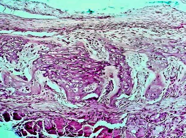
Throughout the entire observation period of up to two months, a dense fibrous capsule around the implant persisted; it did not thin simultaneously with bone remodeling, which could subsequently cause late implant instability.
A group of animals with calcium hydroxyapatite-coated implants. Same as in the uncoated group, no inflammatory response to implantation was observed. HAp coating particles migrated and integrated with peri-implant tissues from the early stages of observation. We did not detect any physiological response to the coating particles. As expected, HAp actively stimulated bone remodeling as early as Week 2 of observation. Neoangiogenesis developed quite actively in the peri-implant zone as well (Figure 2).
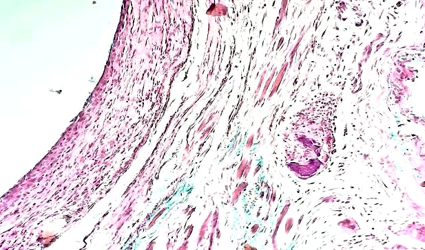
It is worth noting that in this group (similar to the control group), during all observation periods, the formation of a fibrous capsule surrounding the implant was recorded. However, by the later stages of observation, the capsule’s thickness thinned simultaneously with the completion of bone remodeling in the peri-implant zone, which can subsequently prevent late implant migration.
A group of animals with implants covered with a combined calcium hydroxyapatite coating and an antibacterial film (HAp+AB). When studying the morphological picture in this group over time, we detected gelatinous AB particles only up to Week 2 of observation; after that, they were resorbed. Characteristically, these coating particles, as well as the HAp particles, did not provoke any noticeable cellular response in the rat body (Figure 3).

Contrary to the HAp implant group, we did not observe pronounced thinning of the formed fibrous capsule by Month 2 of observation, and bone remodeling processes were still occurring.
A group of animals with chitosan-coated implants. When analyzing microscopic specimens from this group, the following should be noted. As in all groups over time, we did not observe any inflammatory response in the peri-implant zone. Chitosan particles were detected primarily during Week 1 after implantation. By Week 2, they were almost completely resorbed, and there was no cellular response to them. It is worth noting distinct stimulation of osteogenesis in this group, which was particularly pronounced by the end of Month 1 of observation (Figure 4); whereas by Week 2, the intensity of bone remodeling was significantly lower than in the HAp group and in the group with combined HAp+AB coating. The thinning of the formed fibrous capsule by the end of the observation period, as well as the gradual completion of bone remodeling processes, were very important because they ensured late implant stability.

Discussion
All of the studied implant types are popular in medicine. Titanium (Ti) and its alloys are widely used as metallic bone implants due to their remarkable mechanical properties and biocompatibility [13]. In 2024, N.N. Didenko et al. demonstrated on in vivo experimental models that the osseointegration properties of the material were improved by electron beam irradiation processing of a titanium alloy coated with 25 nm ALD TiO2, as well as with an ion-plasma TiO2 coating [14].
Chitosan is a biocompatible, biodegradable polysaccharide derived from chitin. It is widely used in implantology to improve implant biocompatibility and stimulate tissue regeneration [15].
Since HAp is the main mineral component of bones and teeth, it is effectively used in medicine as an implant to replace damaged bone, as well as a coating or filler [16]. HAp-based implant materials often have a porous, interconnected architecture that mimics the intracellular matrix, thereby promoting cell proliferation and tissue regeneration. Researchers at the Institute of Mathematical Problems of Biology––the Branch of Keldysh Institute of Applied Mathematics (Russian Academy of Sciences), conducted high-precision calculations of the effect of substituting magnesium for calcium in HAp [17].
Tomsk scientists studied the in vitro and in vivo biological activity of copper-modified HAp implants but found high cytotoxicity of this material [16].
We demonstrated the biological compatibility of all implant samples with different coatings. Moreover, the duration of the inflammatory response of the wounds in animals with implants covered with a combined coating (HAp+AB) was the shortest, averaging 2.5±0.2 days (p=0.044). When studying the morphological picture in this group over time, in addition to the well-known role of HAp, we were interested in the new coating containing an antibacterial agent, which was resorbed by the end of Week 2 of observation and ensured more rapid resolution of inflammation in the postoperative wound area. This is largely consistent with our concept of prolonged action of the antibacterial agent in the peri-implant zone. We did not observe any noticeable cellular response in the rat body, indicating good biocompatibility of the implant.
Various antibacterial implant coating technologies effectively address infection issues, contributing to a reduction in the incidence of postoperative infection by up to 90%, as demonstrated in both preclinical studies and clinical practice [18].
These results are consistent with our proposed concept of selectively using new coatings and their combinations in the manufacture of novel endoprostheses and other metal structures for reconstructive and regenerative surgery.
Conclusion
Overall, the following common microscopic features can be noted across all groups. In the zone of reactively altered and newly formed bone tissue with active intraosseous osteoblasts, there was no lysis or resorption of preexisting bone tissue in both the cortical and cancellous layers, nor was there a periosteal inflammatory response (no signs of exudative periostitis) in the presence of signs of periosteal bone formation.
All groups demonstrated a consistent maturation of immature bone tissue into lamellar bone tissue, with full morphofunctional characteristics of the developing peri-implant tissue. An increase in the severity of perifocal neoangiogenesis was recorded in the first two weeks. No reactive changes in hematopoiesis were observed, based on the composition of bone marrow elements in the intertrabecular spaces in the adjacent area, indicating full biocompatibility of the implant materials. Just a few observations with mild degenerative changes in adjacent soft tissues (muscle, fat) were identified, primarily in the control group with uncoated samples.
In the groups with coated implants, inclusion of coating particles into the forming fibrous capsule was observed, thereby demonstrating their deeper penetration over time, with a decrease in inclusion size and their disappearance, biodegradation, and objective signs of angiogenesis stimulation, fibroblast proliferation, and osteogenesis without a perifocal inflammatory response.
HAp and chitosan coatings promoted accelerated osseointegration through stimulating angiogenesis, stromal collagen synthesis, and bone formation, with full biocompatibility and rapid biodegradation. These coatings have a pronounced stimulating effect on osteogenesis and neoangiogenesis. When using a combined HAp+AB coating, the effect of HAp on osteogenesis is also present, albeit slightly delayed, possibly due to the resorption time of the antibacterial coating.
Our morphological study showed that the use of all implant types was not accompanied by exudative inflammation, did not cause an allergic reaction (complete absence of eosinophils in the infiltrates), or granuloma formation, nor did it cause severe stromal fibrosis with the proliferation of coarse scar tissue.
Conflict of Interest. None declared by the authors.
Author contributions. All authors contributed equally to the preparation of the manuscript.
Funding. The authors declare no external funding for the study and publication of this article.
Compliance with Bioethics Rules. The study was conducted in accordance with the ethical standards for the treatment of animals adopted by the European Convention for the Protection of Vertebrate Animals used for Experimental and Other Scientific Purposes.
Korytkin AA, Orlinskaya NYu, Novikova YaS, et al. Biocompatibility and osseointegration of titanium implants of varying porosity with and without a calcium phosphate coating. Current Technologies in Medicine 2021; 13(2): 52. (In Russ.) https://www.doi.org/10.17691/stm2021.13.2.06
Kędzia O, Lubas M, Dudek A. Bioactive coatings and the safety of using metal implants. System Safety: Human – Technical Facility – Environment 2024;6(1):227-38. https://www.doi.org/10.2478/czoto-2024-0025
Yadav S, Nath A, Suresh R, et al. Advances in implant surface technology: A biological perspective. Cureus 2025; 17(1): e78264. https://www.doi.org/10.7759/cureus.78264
Bohara S, Suthakorn J. Surface coating of orthopedic implant to enhance the osseointegration and reduction of bacterial colonization: A review. Biomater Res. 2022; 26(1): 26. https://www.doi.org/10.1186/s40824-022-00269-3
Joshi MU, Kulkarni ShP, Choppadandi M, et al. Current state of art smart coatings for orthopedic implants: A comprehensive review. Smart Materials in Medicine 2023; 4: 661-79. https://www.doi.org/10.1016/j.smaim.2023.06.005
Sheveleva VS, Peleshok SA, Titova MV, Kondratenko AA. A review of experimental models for studying the effectiveness and safety of titanium implants. Laboratory Animals for Scientific Research 2024; (3): 78-89. (In Russ) https://www.doi.org/10.57034/2618723X-2024-03-07
Bugaev GA, Antoniadi YuV, Pomogaeva EV, et al. Current understanding of using porous titanium implants and its alloys for bone defect replacement. Polytrauma 2023; (2): 94-102. (In Russ.) https://www.doi.org/10.24412/1819-1495-2023-2-94-102
Stogov MV, Emanov AA, Kuznetsov VP, et al. Comparative evaluation of osseointegration of new percutaneous implants made of ultra-fine-grained Ti Grade 4 alloy. Genius of Orthopedics 2023; 29(5): 526-34. (In Russ) https://www.doi.org/10.18019/1028- 4427-2023-29-5-526-534
Burkhanbayeva T, Ukhov A, Fedorishin D, et al. Development of new composite materials by modifying the surface of porous hydroxyapatite using cucurbit[n]urils. Materials 2024; 17(9): 2041. https://www.doi.org/10.3390/ma17092041
Xionggang Ch, Jianhong Zh, Yu Q, Ling Zhou Zh. Antibacterial coatings on orthopedic implants. Materials Today Bio 2023; 19: 100586. https://www.doi.org/10.1016/j.mtbio.2023.100586
Mukhametov UF, Lyulin SV, Borzunov DYu. Potential for the use of hydroxyapatite-based bone grafting materials in spinal surgery. Creative Surgery and Oncology. 2022; 12(4): 337-44. (In Russ.) https://www.doi.org/10.24060/2076-3093-2022-12-4-337-344
Wakabayashi H, Hasegawa M, Naito Y, et al. Long-term outcome of treating periprosthetic hip joint infection with local antibiotics delivered through antibiotic-impregnated calcium hydroxyapatite. J Clin Med. 2024; 13(23): 7469. https://www.doi.org/10.3390/ jcm13237469
Jia R, Li K, Li J, et al. Surface modification of hydroxyapatite coating for enhanced antibiotic therapy. Coatings 2024; 14(4): 477. https://www.doi.org/10.3390/coatings14040477
Didenko NN, Bobryshev DV, Dolgalev AlAl, et al. Biocompatibility of cell cultures with new titanium alloy samples for dental implantology. Medical Bulletin of the North Caucasus 2024; 19(3): 260-2. (In Russ.) https://www.doi.org/10.14300/mnnc.2024.19059
Sidorova SV, Moiseev KM, Vasiliev DD, et al. Plasma treatment of material surfaces for photonics applications. FRos. 2022; 16(4): 288-95. (In Russ.) https://www.doi.org/10.22184/1993-7296
Bystrov VS, Paramonova EV, Avakyan LA, et al. Effect of magnesium substitution on structural features and properties of hydroxyapatite. Materials 2023; 16(17): 5945. https://www.doi.org/10.3390/ma16175945
Schneidmiller AA, Fedorishin DA, Korotchenko NM. Study of the biological activity of copper-modified hydroxyapatite samples. Transactions of the Kola Science Center of the Russian Academy of Sciences. Chemistry and Materials Science 2021; 11(2): 285-90. (In Russ.) https://www.doi.org/10.37614/2307-5252.2021.2.5.057
Romanò CL, Bozhkova SA, Artyukh V, et al. The use of implants with antibacterial coating in orthopedics and traumatology: The current state of the problem. Traumatology and Orthopedics in Russia 2019; 4(25): 64-74. (In Russ) https://www.doi.org/10.21823/2311-2905-2019-25-4-64-74